近期,密歇根大學Marco C. Bottino教授在科愛創辦的期刊Bioactive Materials發表研究論文。該研究報道了具有組織特異性的三維熔體電書寫支架,指導人牙周膜干細胞(hPDLSCs)的分化并介導巨噬細胞極化。在500 μm鏈間距支架上的hPDLSCs,在21天內表現出較高的成骨表達,而隨機定向纖維支架上的細胞表現出M1標記的上調。在原位下頜開孔缺損模型中,研究結果顯示,組織特異性支架)可以增強對天然牙周組織再生的模擬。
引言
牙周炎是一種非常普遍的慢性炎癥,目前治療牙周炎的方法包括徹底的齦下刮除、根面平整和翻瓣手術,并通過可降解膜引導組織再生。總的來說,盡管有證據表明,在上述治療后可能會出現一定程度的牙周再生,但由于牙周缺損的多組織復雜性和三維性質,在臨床中,模擬原生牙周組織的能力可能是不切實際的。
眾所周知,牙周再生需要同時處理軟(PDL)和硬組織界面。重要的是,獲得定向良好的PDL纖維與牙槽骨再生是至關重要的。增材制造(AM)是一個快速發展的領域,已在再生口腔醫學中得到應用。熔體電寫入(MEW)是一種替代AM工藝,可以生產具有明確宏觀和微觀結構特征的纖維支架,如孔隙率(即鏈間距)、纖維排列和直徑。盡管氟化磷酸鈣(F/CaP)涂層的MEW聚合物纖維支架在支持PDL新生生物的同時能夠實現大量的牙槽骨再生,但最近的文獻表明,單相支架策略在骨-韌帶界面重建PDL角結構的能力有限。這些結果進一步強調了個性化解決方案的需求,可以指導牙周附著裝置的協調生長和發育。
近年來,支架介導的策略提供三維模板和仿生細胞外基質微環境,同時指導人體的免疫反應已被建議在組織工程應用。先天性免疫反應在生物材料植入后發揮重要作用,以減輕促炎反應,并允許巨噬細胞極化。促炎M1巨噬細胞轉化為抗炎巨噬細胞(M2)表型對生物材料植入部位的整體炎癥反應至關重要。
因此,對于目前使用的可溶性生物分子來說,生物材料介導支架即使不是一種可能的替代品,可以指導祖細胞的反應,以促進組織愈合并最終再生。在這項研究中,我們利用MEW在確定的纖維方向(隨機定向或對齊支架)和鏈間距(即,纖維的交叉鋸齒圖案,鏈間距為500 μm和250 μm;下文簡稱為高度有序的腳手架)。體外研究了纖維取向和鏈間距對人源性牙周韌帶干細胞(hPDLSCs)附著/增殖和分化的影響,以確定用于牙周重建的組織特異性支架中最有利的PDL特異性區域。總的來說,我們探索了一種多功能和創新的平臺(即MEW),最終可能能夠指導組織特異性(PDL的對齊纖維和牙槽骨高度有序的500 μm鏈間距氟化磷酸鈣[F/CaP]涂層纖維)干細胞分化和巨噬細胞極化的帶狀智能生物材料支架的發展,作為有效牙周組織再生的潛在個性化治療。
1. MEW PCL支架的制備與表征
MEW成功地用于制造具有不同設計的3D纖維支架,即高有序(即交叉0/ 90°,250 μm(小)和500 μm(大)的鏈間距,以及高定向(對齊)和無定向(隨機)的纖維配置(圖1))。獲得的支架具有高多孔性和高選擇性呈光滑纖維形態。在收集板上任意沉積PCL纖維,就會產生無定向的隨機纖維支架。各種MEW PCL支架的代表性掃描電鏡圖像顯示熔體電印跡聚合物(即聚ε-內酯)支架具有組織特異性屬性,如纖維形態(隨機vs對齊)和高度有序(0?/90?交叉圖案)結構,具有明顯的鏈間距(小250 μm和大500 μm)。總體而言,制備的支架的纖維直徑(μm)分別為2.3±0.1、2.5±0.2、2.6±0.3和4.2±0.7,在排列、大、小股間距和隨機沉積的纖維中,孔隙率分別為52.9%、91.7%、85.5和30.9%。
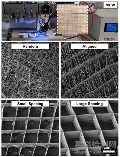
Fig. 1. Melt electrowriting (MEW) setup to fabricatefibrous scaffolds of distinct fiber configuration andhighly-ordered architectures. Representative SEMimages of the various MEW PCL scaffolds show meltelectrowritten polymeric (i.e., poly(ε-caprolactone)scaffolds with tissue-specific attributes such as fibermorphology (random vs. aligned) and highly-ordered(0?/90? crosshatch pattern) architecture with distinctstrand spacings (small 250 μm and large 500 μm).Overall, the as-produced scaffolds display fiber diameters (in μm) ranging from 2.3 ± 0.1, 2.5 ± 0.2,2.6 ± 0.3 and 4.2 ± 0.7 and porosity of 52.9%,91.7%, 85.5 and 30.9% in aligned, large and smallstrand spacing to randomly-deposited fibers,respectively.
2. hDPLSCs細胞的排列和分化取決于纖維取向和支架結構
纖維組織在組織界面的特定排列在優化生物力學和生物物理反應方面具有重要作用。因此,本研究探索了一種組織特異性引導支架,通過在結構上和成分上量身定制的帶狀支架(即用于PDL區的高度定向(排列)的纖維和用于骨區的高度有序的(500μm鏈間距)、納米結構的氟化帽涂層多孔性支架)的新制造來鼓勵牙齒-韌帶-骨界面的同時再生。
在我們的研究中,hPDLSCs在增殖和分化能力方面對不同的支架結構和纖維配置做出了反應(圖2)。而種植在無取向(隨機)和高度取向(取向)纖維上的hPDLSCs與具有可變股間距的MEW PCL支架相比顯示出更高的增殖率(圖2A),在整個體外培養期間,與大鏈間距相比,小鏈間距的增殖率明顯更高(圖2A),這是由于更高的表面積和更小的鏈間距(孔徑),增強了初始細胞附著。
Fig. 2. Attachment and proliferation of hPDLSCs onMEW PCL scaffolds with aligned and randomly oriented fiber configurations, and 250 μm and 500 μmstrand spacings. (A) Cell viability of hPDLSCs seededon the scaffolds using AlamarBlue assay over 7 days.(B) Representative SEM images of hPDLSCs proliferation on the scaffolds after 3 and 7 days. Note thecharacteristic cell stretch along the fibrous walls (AllSEM images have the same scale bar). A more pronounced spreading was detected along the scaffoldswith randomly oriented fibers (white arrows indicatethe direction of filopodia protrusion). (Mean ± SD, n= 3). *p < 0.05, **p < 0.01, ***p < 0.001.
通過SEM(圖2B)和熒光(phalloidin染色)成像(圖3),可以明顯地看到與各種MEW PCL支架誘導的接觸引導相關的hPDLSCs形態的直接變化。具有平行沉積纖維的高定向(對齊)支架允許hPDLSCs平行于纖維的長軸方向對齊(圖2B),而在隨機和高度有序的支架(即小和大鏈間距)中,hPDLSCs表現出多邊形形狀,細胞骨架多方向延伸,取向混亂。因此,hPDLSCs在對齊的纖維上培養時采用了典型的紡錘形形態,這是以前在人類成纖維細胞中觀察到的特征。
Fig. 3. Patterns of hPDLSCs alignment on the various MEW PCL scaffolds. (A) Representative CLSM images show a variable pattern of cells’ bridging, following thefibers’ arrangements at day 3 and 7. DAPI (blue) and phalloidin (red) fluorescent staining (scale bar = 50 μm). (B) Histograms of PDLSCs nucleus angulation onscaffolds with aligned and randomly oriented fiber configurations as well as crosshatch 0/90? arrangement and 250 μm and 500 μm strand spacings corresponding toconfocal images.
為了評估hPDLSCs在工程支架上的分化潛力,在缺乏分化因子的情況下,評估了特異性編碼韌帶生成(骨膜蛋白[POSTN]、鞏膜[SXC]和膠原III [COL3])和成骨(runt相關轉錄因子2 [RUNX2])的基因表達(圖4)。在第3、7和14天,所選的韌帶生成基因(即骨膜蛋白、鞏膜和Col3)的mRNA水平在纖維對齊的支架中顯著升高,而成骨標記物(Runx2)在橫線0/90?250 μm和500 μm鏈間距的支架中顯著升高。
Fig. 4. Ligamentogenic and osteogenic differentiation of hPDLSCs seeded on the various MEW PCL scaffolds. The mRNA levels at days 3, 7, and 14 of selectedligamentogenic genes (i.e., periostin, Scleraxis, and Col3) were significantly higher in scaffolds with aligned fibers, whereas an osteogenic marker (Runx2) wassignificantly higher in crosshatch 0/90? scaffolds with 250 μm and 500 μm strand spacing. (Mean ± SD, n = 4). *p < 0.05, **p < 0.01.
3. 巨噬細胞的伸長和極化取決于纖維取向和支架結構
研究通過控制支架結構(鏈間距)和纖維結構(對齊vs隨機),將RAW 264.7細胞(巨噬細胞)接種在確定的支架上,在培養基中加入的炎癥標志物表達誘導物——大腸桿菌脂多糖(LPS),通過qPCR和ELISA檢測巨噬細胞表型標志物的表達變化。在隨機定向的纖維支架上培養的RAW 264.7細胞能夠在第1天保持圓形表型,而當巨噬細胞在對齊的支架上培養時,細胞伸長更為突出(圖5)。同時,在所有測試支架上,未受到刺激和LPS刺激的RAW 264.7細胞的細胞擴散和伸長保持相當(圖5)。值得注意的是,即使在炎癥刺激的環境中,纖維排列和鏈間距傾向于影響巨噬細胞伸長,這被認為是促進表型標記物從M1到M2表達的變化。
Fig. 5. Representative SEM images showing RAW264.7 cells’ morphology of spontaneously differentiated and LPS-stimulated macrophages. MEW scaffoldswith varying fiber orientation and architecture(strand spacing), namely, random, aligned, and scaffolds of 250 μm and 500 μm strands spacing at 0?/90?-oriented junctions. The images show mixed patterns of macrophages spreading, the round shapetypical for M1 and more pronounced elongatedpattern typical for M2 phenotype.
LPS刺激巨噬細胞在不同纖維取向和結構(鏈間距)支架上的細胞因子釋放。使用在不同支架上培養巨噬細胞后提取的上清液測量釋放的細胞因子,并將其與對照TCP進行7天的比較(圖6)。
Fig. 6. Cytokine release of LPS-stimulated macrophages on scaffolds with different fiber orientation and architecture (strand spacing). Released cytokines weremeasured using supernatants extracted after culturing macrophages on different scaffolds, and they were compared to control TCP over 7 days. (Mean ± SD, n = 3).*p < 0.05; **p < 0.01, ***p < 0.001.
此外,在lps刺激的巨噬細胞中評估了7天的基因表達譜(圖7)。qPCR檢測M1、IL-1β、IL-6和M2、IL-10 7 d內特異性標記物的表達。我們認為M2表型與M1表型具有不同的功能,通過刺激細胞增殖和ECM沉積上調修復和再生相關因子,以及血管生成效應。總之,我們的數據表明,排列的纖維結構和具有大小鏈間距的高度有序的支架設計促進巨噬細胞向M2表型極化。
Fig. 7. Gene expression profile of LPS-stimulated macrophages, the level of IL-10, MRC1 and IL-1β. Expression of specific markers was measured using qPCR for M1,IL-1β and IL-6 and for M2, IL-10 over 7 days; data was compared to TCP as a reference sample. (Mean ± SD, n = 3). *p < 0.05; **p < 0.01, ***p < 0.001.
4. 組織特異性支架指導牙周再生的體內評估
為了闡明組織特異性屬性的重要性,我們詳細制作了一種新型納米結構氟化磷酸鈣(F/CaP) MEW PCL支架。所制備的組織特異性支架具有明顯的纖維取向(圖8A)。特別是,SEM分析表明,設計的PDL區域在~200 μm高度與原始PDL空間相似,而骨區域在~800 μm高度高度具有高度有序(0/90?交叉鋸齒)和多孔(500 μm鏈間距)。FTIR光譜數據顯示(圖8B),F/ cap涂層支架中~565 cm和~960 cm處與磷酸鹽相關的化學官能團,而灌注膠原蛋白的支架和膠原蛋白也顯示了酰胺基團,證實了膠原蛋白在MEW支架內的成功滲透。掃描電鏡(SEM)和紅外光譜(FTIR)分析表明,在不改變所使用的基底材料成分的情況下,可以有效地制備膠原灌注支架。

Fig. 8. Fabrication and characterization of tissue-specific zonal scaffolds. (A) Representative SEM images for tissue-specific scaffold with aligned PDL zone and 500μm strands spacing for bone zone, and SEM images for collagen-infused scaffolds. (B) FTIR spectra of collagen, F/CaP-coated MEW PCL scaffold, and F/CaP-coatedMEW PCL scaffold infused with collagen. Data show chemical functional groups related to phosphate at ~565 cm− 1 and ~960 cm− 1 in F/CaP-coated scaffolds, whilecollagen-infused scaffolds and collagen also show the amide groups, confirming successful permeation of collagen within the MEW scaffolds. (*) indicates thepresence of PCL. (C) Generation of the rat mandibular periodontal fenestration model. Photographs of a rat mandible after the incision, flap elevation, creation of thedefect, and implantation of tissue-specific scaffold in the defect region.
利用已建立的大鼠下頜骨牙周開窗缺損模型,對工程纖維支架支持新組織沉積的潛力進行評估(圖8C)。根據我們的組織學和免疫組化結果,制備的帶式支架的成分(即F/ CaP涂層)和結構組織(纖維排列)導致牙槽骨和PDL同時再生(圖9)。microCT分析顯示,在所研究的支架組之間,缺損部位的骨形成存在顯著差異(圖9A)。術后3周和6周,不同組骨容量、骨填充和組織礦物密度的μCT評估。與對照組和非包膜組相比,包膜(F/CaP)組和膠原蛋白的存在進一步顯示了骨體積和骨填充的顯著差異(圖9B)。

Fig. 9. MicroCT assessment of bone formation. (A) Representative microCT images of the fenestration defect exposing the distal root of the second molar at 3 and 6weeks in the Control (Sham), Collagen, and Biphasic scaffold with Aligned (PDL) compartment and non-coated and coated (F/CaP) and aligned and coated (F/CaP) +collagen. Transverse views highlight the visual differences between the area and density of bone regenerated within the defect. (Scale bar = 1 mm). (B) μCT assessments of bone volume, bone fill, and tissue mineral density at 3 and 6 weeks after surgery, within the different groups. The coated (F/CaP) group and the presenceof collagen further show significant differences for bone volume and bone fill compared to both the control and non-coated groups. Mean ± SD (n = 6). ANOVA: *p< 0.05; **p < 0.01.
對新形成的牙周組織的組織學評估顯示,所提出的組織特異性分區支架(即對齊/ F-CaP涂層支架)在再生能力(圖10)。對照組由于上皮組織向下生長,表現為軟組織侵犯;單用膠原蛋白均不能促進牙周硬組織和軟組織的再生。值得注意的是,我們的組織特異性分區支架為組織形成提供了指導,與假組(對照組)和膠原組相比,對齊/ F-CaP涂層支架的新骨形成量更高。有趣的是,兩種組織特異性分區支架(輸注和不輸注膠原蛋白)都維持了與天然牙周組織相似的PDL空間。總體而言,F/ cap涂層的MEW PCL支架與未涂層(NaOH蝕刻)和原始支架相比,具有更高的抗拉強度和楊氏模量。未來的研究不僅要解決再生組織的生物力學問題,而且要解決循環(咀嚼)負荷下支架性能和整體完整性的問題。
Fig. 10. Masson’s trichrome staining of periodontal defects treated with the distinct scaffolds and evaluated after 6 weeks post-scaffolds implantation. Representative Masson’s trichrome-stained horizontal cross-section of Control (Sham), Collagen, and tissue specific scaffolds with aligned fiber configuration (PDL)compartment and coated (F/CaP) fibers (bone compartment) and tissue specific scaffold infused with collagen at 6-weeks post-implantation. Yellow dashed linehighlights the area of scaffold placement and new bone formation. White arrowhead for PDL, NB: new bone; R: Root surface; St: Soft tissue.
10. 結論
總之,我們的研究結果為支架結構(鏈間距)、纖維結構和成分(F/CaP涂層)的形態和功能變化提供了證據。具體而言,排列整齊的纖維強烈支持韌帶形成,而排列整齊的支架結構(束間距為500 μm)支持成骨,這在牙周組織工程中尚未見報道。此外,纖維方向和鏈間距被發現與巨噬細胞的形態伸長和向促愈合的M2表型分化直接相關。體內研究結果證實,結構定制的支架具有高度定向(對齊)的MEW PCL纖維形成的PDL區和由F/cap涂層的MEW PCL纖維組成的骨區,其鏈間距為500 μm,可以在一個成熟的嚙齒動物下頜開窗缺損模型中實現協調的牙周組織再生。總之,我們的研究結果表明,MEW最終可能導致區域性生物材料介導支架的發展,能夠指導組織特異性干細胞分化和巨噬細胞極化。這種方法可以作為一種個性化的治療方法,用于炎癥驅動疾病(如牙周炎)的有效組織再生。
原文信息: Arwa Daghrery, Jessica A. Ferreira, Jinping Xu, Nasim Golafshan, Darnell Kaigler, Sarit B. Bhaduri, Jos Malda, Miguel Castilho, Marco C. Bottino.
Tissue-specific melt electrowritten polymeric scaffolds for coordinated regeneration of soft and hard periodontal tissues.
Bioactive Materials, 19 (2023) 268–281.